3D Geometry SDC (Second Dose Check) for Cyberknife with RayStation DICOM Export
Authors:
Ivy Au1, Deirdre Hutton1, Hidayat Ghani2, Carlos Bohorquez2, and Ian M Hanson1
1Auckland Radiation Oncology, Epsom, Auckland, New Zealand
2LifeLine Software, Tyler, Texas, United States of America
INTRODUCTION
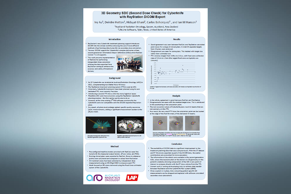
RayStation’s new Cyberknife treatment planning support introduces DICOM into the clinical workflow allowing the use of more efficient methods of performing robust plan QA via secondary dose calculation. The new DICOM support for Cyberknife’s complex deliveries utilizes second generation Information Object Definitions (IODs) which RadCalc version 7.3.2.0 supports. This work presents implementation of RadCalc for performing independent dose calculation utilizing the data exported from RayStation aiming to enhance the accuracy and safety of treatment delivery.
BACKGROUND
An S7 Cyberknife was installed at Auckland Radiation Oncology (ARO) in 2022, complementing two Elekta Versa HD linacs. The RayStation treatment planning system (TPS) is used at ARO. Historically, Cyberknife treatments have been planned using its own dedicated TPS, most recently Precision. Introducing a second TPS into a clinic has many logistical issues. Therefore ARO went live exclusively using the RayStation Cyberknife planning module – the first centre world wide to do so. However, at the time existing 2nd MU software solutions for the Cyberknife were not compatible with the DICOM exported Raystation plans. As a result, all plans must undergo patient specific quality assurance (pQA) measurements, adding a significant measurement burden to the physics team.
METHOD
Pre-configured machine models provided with RadCalc were fine tuned with the measured output factors, off axis ratios, and PDDs. Existing clinical plans were exported to RadCalc. Doses to reference points were calculated and compared to values from RayStation. All treatment plans had been validated by independent dose measurement using an SRS MapCHECK to measure each PTV. Work focused on SRS plans delivered using the Fixed Cone collimator system of the Cyberknife.
RESULTS
Good agreement was seen between RadCalc and RayStation calculated point doses for a range of clinical plans. In total 35 separate targets from 23 plans were analysed. Disagreement was seen for some plans. This tracked with target size rather than collimator size. PTV volumes ranged from 0.04cc to 2.01cc, with fixed cone collimator sizes of 0.5cm to 1.5cm (the largest fixed cone we typically use clinically).
ANALYSIS
In the whole, agreement is good between RadCalc and RayStation. Disagreements are seen with the smallest target sizes. This is attributed to the positioning of the calculation point. The non-isocentric nature of Cyberknife plans result in beams that are not centred w.r.t the PTV. As a result, for very small PTV sizes the calculation point can be located at the edge of the field for many of the delivered CK beams.
CONCLUSIONS
The availability of DICOM data is a significant improvement to the treatment planning data that was used previously. With the CT dataset and RT Structures exported as part of the DICOM data, the scatter contributions are accounted for more accurately. The information of the robotic arm available in the second generation IODs, allows the reconstruction of the delivery in 3D geometry on the patient’s anatomy allowing computation of the SDC to any point of interest to compare directly with the RT dose volume from the TPS. Current work is looking at developing tools to validate data transfer between RayStation and the CyberKnife R&V system (iDMS). Once a system is in place, time consuming patient specific QA measurements can be dropped and replaced with software calculated secondary dose calculations.

